JD’s laboratory has another fantastic post for you to read! This particular post is addressing one of the most common skin cancers, squamous cell carcinoma. Now this one tends to be more aggressive than the one from the previous post, basal cell carcinoma.
There are cells in the epidermis called squamous cells and they are responsible for producing the keratotic barrier between your skin and the outside world. In the malignant form, these cells can travel down into the skin and in some cases develop what we call “keratotic pearls.” The squamous cells even produce keratin to the point they grow outward from the skin to form “horns.”
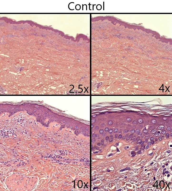
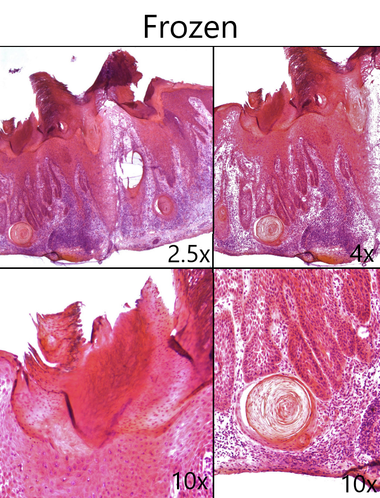
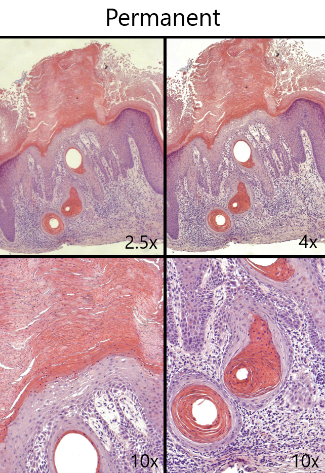
The first image shows one of the ways squamous cell carcinoma can appear on the skin. The second image is to show what normal skin is supposed to look like under the microscope. The third set of images is what we call a “frozen sections.” It’s a biopsy of a suspected skin cancer that is tested in the lab in under an hour for fast results so the providers can discuss treatment options before you ever leave the clinic! That’s one of the things that sets JD apart from other clinics in the area. The fourth set of images is the same biopsy but stained with our much slower stain for “permanent sections” which provides clearer imaging
The pink dense material seen at the top and in the circular structures (the pearls mentioned earlier) is the keratin produced by the squamous cells which looks drastically different from the strand-like keratin seen in the control sample. The light purple cells surrounding the pearls and at the base of the large pink growth are the malignant squamous cells.